
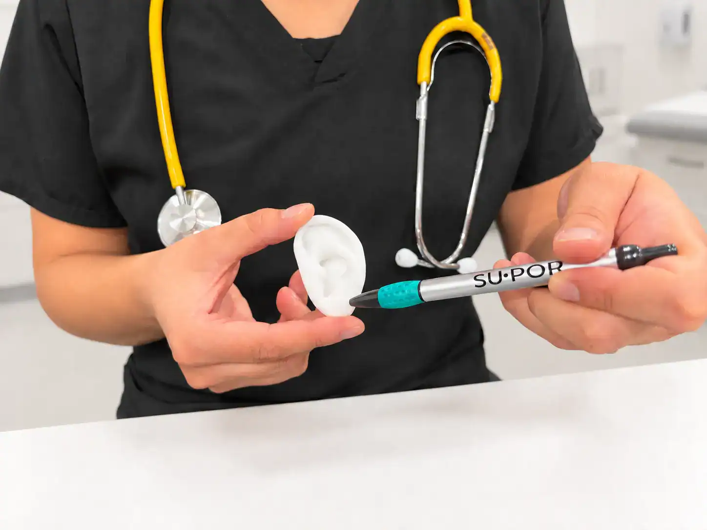
Microtia is a congenital malformation of the external ear that presents significant challenges in reconstructive surgery worldwide. The severity of the condition ranges from mild structural abnormalities to anotia, the complete absence of the external ear.1
The most common technique to reconstruct the ear involves using autologous costal cartilage grafts harvested from the patient’s rib cartilage.1 An estimated 91.3% of surgeons use this technique on patients ranging from ages six and up using a three-stage process: implantation of a tissue expander, framework fabrication using harvested rib cartilage, and implantation in the pocket. This is followed by tragus and concha reconstruction.1
In some procedures, a fourth stage is performed, where the tragal reconstruction and concha excavation is conducted with simultaneous contralateral otoplasty utilizing an additional piece of harvested cartilage to ensure the ear is elevated.4
Porous polyethylene implants offer a viable alternative to conventional autologous cartilage grafts. Porous polyethylene is known for their lower complication rates, ease of use, and overall impact. This offers surgeons greater flexibility while making procedures much easier on pediatric patients.2
Autologous Costal Cartilage Grafts vs. Porous Polyethylene Implants
While common, microtia reconstruction is a relatively rare procedure for younger pediatric patients; some surgeons advocate for delaying the procedure until a patient is at least 10 years old to avoid potential risks of chest-wall deformities or pneumothorax.1,2
Despite delaying surgeries, the procedure has a 25% overall complication rate. Patients waiting until they’re older may also subject them to bullying due to their pre-operative physical appearance.2
Porous polyethylene implants have been shown across multiple peer-reviewed studies to deliver better aesthetic results, require fewer procedures, and have shorter surgery and recovery times. In addition, they have been shown to be suitable for patients as young as age three.2
Unlike traditional surgeries that rely on developed rib cartilage for harvesting, the use of porous polyethylene implants eliminates the need for these invasive procedures. Porous polyethylene auricular reconstruction avoids scarring, risk of chest-wall deformities or pneumothorax, along with multi-step procedures, and overnight stays.2 Procedures can be done in a single-step process with a shorter recovery period. The technique involves the placement of a temporoparietal flap over the implant for soft tissue coverage, which is typically a one or two stage procedure.2
Complications with porous polyethylene implants are also significantly lower compared to traditional microtia procedures, including a 7% exposure, and a 2.7% fracture risk.2 Procedures are also less complex.
The use of porous polyethylene implants is ideally suited for microtia surgeries because of its high success rate.3 SuᐧPor implants offer an even lower risk of complications to patients, with all of the proven benefits of porous polyethylene and more.
In a study of 649 children who underwent microtia reconstruction, patients who had autologous rib cartilage procedures were significantly more likely to undergo additional procedures at 90 days, 180 days, and one year, when compared to patients who had reconstruction surgeries using porous polyethylene implants.3 The study further found that 67% of patients who had traditional microtia surgeries had another procedure within one year; procedures dropped to 20% for patients using implants.3
Less Complications and More Successful Healing with SuᐧPor
SuᐧPor’s porous polyethylene materials are fully biocompatible, integrating with existing tissue with minimal issues. Unlike other porous polyethylene implants, SuᐧPor has demonstrated less than half of 1% of overall complications, including breakage, rejection, or infection. Leading surgeons have described SuᐧPor as the superior implant option, reporting zero exposure issues across multiple craniomaxillofacial procedures.5
They’re also uniquely ideal for pediatric patients. Ears grow to about 85% of adult sizes by age four, allowing for implant use much earlier than autologous rib cartilage procedures.2 One-piece ear implants are available in multiple shapes and sizes to suit most patient needs.
SuᐧPor ear implants can be contoured, trimmed, drilled, sutured, and securely fixated in the OR and in vivo without the need for specialized additional tools. The porous polyethylene ear implants create a living graft that allows for fibrovascular in-growth, resulting in short and long-term stabilization and infection resistance.
For cases requiring patient-centric solution, SuᐧPor Patient-Specific Implants offer a customized fit. Designed and manufactured based on the patient’s individual anatomy, using CT data or high fidelity laser scans to support the design process, SuᐧPor PSI implants are precisely design to help surgeons provide the best care for their patients.
Like all Su·Por offerings, Su·Por auricular implants are designed, produced, and batch-tested for bacterial endotoxin levels in-house. All implants are shipped sterile, with post-market surveillance demonstrating an infection rate of under 0.5%. The implants are MR-safe and radiolucent, supporting future imaging needs post ear-reconstruction.
Explore Su·Por ear implants for pediatric microtia surgery at https://poriferous.com/facial-implants/.
Sources
- Tripathee, S., Xiong M., et al. “Microtia Ear Reconstruction Using Tissue Expander and Autologous Costal Cartilage: Our Experience and Comparing Two Age Groups”. World J Plast Surg. 2019 Sep;8(3):324–330. doi: 10.29252/wjps.8.3.324
- Ali, K., Trost, J., et al. “Total Ear Reconstruction Using Porous Polyethylene”. Semin Plast Surg. 2017 Aug 9;31(3):161–172. doi: 10.1055/s-0037-1604261
- Varagur, K., Zubovic, M., et al. “Porous polyethylene versus autologous costochondral reconstruction for microtia: incidence and analysis of secondary procedures”. Cleft Palate Craniofac J. 2022 Oct 11;61(3):365–372. doi: 10.1177/10556656221132034
- Olshinka, Asaf, Matthew Louis, and Tuan A. Truong. “Autologous Ear Reconstruction.” Seminars in Plastic Surgery 31, no. 3 (2017): 146–51. doi: 10.1055/s-0037-1603959
- Mraček, J., Siedl, M., et al. “Three‐dimensional personalized porous polyethylene cranioplasty in patients at increased risk of surgical site infection.”Acta Neurochirurgica (2024) 166:383 https://doi.org/10.1007/s00701-024-06281-x